Imagine your friend, mid-soccer game, suddenly clutching their groin. They’re in pain, and you’re worried. Could it be a hernia? Knowing how doctors check for a hernia is important. This post explores the process step by step, from initial symptoms to diagnosis. You’ll gain a clear picture of what to expect, reduce anxiety, and be prepared if you or someone you care about experiences similar symptoms.
Key Takeaways
- Doctors use a physical examination to check for a hernia.
- They will ask about your symptoms and medical history.
- Imaging tests like ultrasound may be used for diagnosis.
- A hernia can be identified by a bulge in the affected area.
- The doctor will assess the size and severity of the hernia.
- You will learn about the different types of hernias and their causes.
The Initial Assessment: Unveiling the Clues
The journey to diagnosing a hernia often starts with a conversation. When someone visits a doctor with potential hernia symptoms, the initial assessment involves gathering information and observing the physical presentation. This critical first step helps to narrow down the possible causes of the symptoms and decide if further testing is needed. Accurate information from the patient is important.
The doctor will start by asking about the patient’s symptoms. Common signs include pain or discomfort in the groin, abdomen, or upper thigh. Patients often describe a noticeable bulge. Some people feel a heavy sensation or pressure in the affected area, especially when straining, lifting, or coughing. Questions about the nature of the pain—whether it is sharp, dull, constant, or intermittent—are useful.
Gathering Medical History: Looking Back
The doctor will then delve into the patient’s medical history. This involves inquiring about any previous surgeries, especially abdominal operations, which can increase the risk of hernias. They’ll also ask about any underlying health conditions, such as chronic cough or constipation, that could contribute to increased abdominal pressure. Family history is also important because some hernias have a genetic component.
Factors like age, gender, and lifestyle are important to the overall evaluation. For example, men are more prone to inguinal hernias. Heavy lifting and strenuous activities can also play a role in hernia development. This information helps the doctor evaluate the probability of a hernia and eliminate other potential causes.
Physical Examination Techniques: The Hands-On Approach
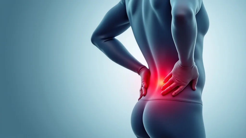
The core of a hernia diagnosis is the physical examination. The doctor will gently feel the affected area to check for a bulge, which is often visible when the patient is standing or straining. They might ask the patient to cough, which increases intra-abdominal pressure and makes the hernia more apparent. The doctor will also try to push the bulge back in, carefully assessing its size and whether it is reducible (can be pushed back). If the hernia is trapped or strangulated, this will also affect the examination.
During a physical examination, the doctor might palpate the area, feeling for any tenderness or changes in the tissue. In the case of an inguinal hernia (a common type affecting the groin), the doctor may examine the scrotum and testicles. In women, the doctor will check the inguinal canal and may need to conduct a pelvic examination to rule out other problems.
Imaging Tests: Seeing Beyond the Surface
While a physical exam often provides enough information to diagnose a hernia, imaging tests play a vital role in confirming the diagnosis, assessing the size and location of the hernia, and ruling out other conditions. These tests provide a more detailed look at the inside of the body. Several different tests can be utilized.
Sometimes, a diagnosis isn’t straightforward, or the physical examination results are unclear. In these cases, imaging tests are essential. These tests are useful when a hernia is suspected, but a bulge isn’t always obvious. Imaging can also help find hernias in people who are overweight, or whose symptoms are not typical. Imaging tests can also help differentiate hernias from other problems that can cause pain or a bulge in the groin or abdomen.
Ultrasound: A Sound View
Ultrasound is commonly used to visualize hernias. This test uses high-frequency sound waves to create images of the abdominal wall and its contents. An ultrasound is painless and doesn’t use radiation. It can help the doctor see the hernia directly, assess its size, and determine if any organs are trapped within the hernia sac. An ultrasound is helpful for detecting hernias that aren’t obvious through a physical examination.
During an ultrasound, the patient lies down while a trained technician, called a sonographer, applies a gel to the area. They then move a small device, called a transducer, over the area to get images. The sonographer can ask the patient to strain or cough during the scan. This can make the hernia more obvious. This helps the doctor to see how the hernia changes under different conditions.
CT Scan and MRI: Detailed Views
Computed tomography (CT) scans and magnetic resonance imaging (MRI) provide detailed cross-sectional images of the abdominal area. These tests can identify a hernia and provide a more comprehensive view of the surrounding tissues and organs. CT scans are particularly useful for detecting hernias, identifying complications, and looking for alternative diagnoses. MRI offers excellent soft tissue detail.
A CT scan uses X-rays to create detailed images of the body. The patient may need to drink a contrast dye to enhance the images. An MRI uses strong magnetic fields and radio waves to create detailed images without radiation. It’s often used when more detail is needed. Both CT scans and MRIs can show the hernia’s size and position and detect any signs of complications, like strangulation. These tests are useful for diagnosing complicated or unusual cases.
Other Imaging Techniques: Exploring All Options
In certain situations, other imaging techniques might be considered. This might include a barium enema, which can help diagnose an internal hernia by highlighting the intestines. It’s rare to use, but can be helpful. The choice of imaging test depends on the type of hernia and the patient’s individual circumstances. The doctor will decide which tests are best for each case.
Sometimes, the doctor might use a laparoscopy, a minimally invasive procedure, to examine the abdominal cavity directly. During a laparoscopy, a small incision is made, and a thin tube with a camera (a laparoscope) is there is a diagnostic uncertainty or when the doctor needs to look more closely at the inside of the abdomen.
Types of Hernias: Understanding the Variations
There are many different types of hernias, each occurring in a different location of the body. They vary in their causes and the specific symptoms they cause. The most common type is an inguinal hernia. Knowing the specific type of hernia is important for deciding on the best course of action. This influences how doctors check for hernia and how they treat the condition.
Understanding the location and characteristics of a hernia is essential for diagnosis. It helps in deciding what treatment is needed. Knowing the type also allows the doctor to provide the patient with accurate information. This also includes the possible risks and benefits of various treatments.
Inguinal Hernias: A Common Challenge
Inguinal hernias are the most frequent type, occurring in the groin area. They develop when a portion of the intestine or other abdominal tissue pushes through a weak spot in the abdominal wall. These hernias are more common in men due to a natural weakness in the inguinal canal. This canal allows the testicles to descend from the abdomen into the scrotum during fetal development.
Symptoms include a bulge in the groin, which may become more obvious when coughing, straining, or standing. Patients may feel pain or discomfort, which may worsen over time. There are two types of inguinal hernias: direct and indirect. Direct hernias usually occur in older adults. Indirect hernias are more common and often result from a congenital weakness.
Femoral Hernias: Affecting the Upper Thigh
Femoral hernias occur in the upper thigh, just below the groin crease. These hernias are more common in women and often appear as a small bulge. They occur when abdominal contents push through the femoral canal, a small channel that carries the femoral artery, vein, and nerves. These can sometimes be mistaken for inguinal hernias.
Femoral hernias tend to be small, but they have a higher risk of becoming strangulated (trapped and having their blood supply cut off). Symptoms may include a painful lump in the upper thigh, and discomfort when standing or walking. Because of the risk of strangulation, femoral hernias usually need surgical repair even if they aren’t causing major problems.
Umbilical Hernias: Around the Belly Button
Umbilical hernias occur near the belly button. They occur when a part of the intestine pushes through a weakness in the abdominal wall. These hernias are common in infants and young children, often resolving on their own as the abdominal muscles strengthen. In adults, umbilical hernias are linked to factors like obesity, multiple pregnancies, or fluid accumulation in the abdomen.
In infants, a small bulge may be noticeable near the belly button, especially when crying or straining. Adult symptoms include a lump near the belly button and may cause pain or discomfort. If the hernia doesn’t go away, or if it grows larger, surgery may be necessary to prevent complications.
Hiatal Hernias: The Esophageal Connection
Hiatal hernias occur when the upper part of the stomach pushes through the diaphragm into the chest. There are two main types: sliding hiatal hernias and paraesophageal hernias. Sliding hiatal hernias are more common, where the stomach slides up into the chest. Paraesophageal hernias are less frequent. The stomach pushes through the diaphragm next to the esophagus.
Symptoms of hiatal hernias include heartburn, acid reflux, chest pain, and difficulty swallowing. The symptoms are often aggravated by lying down, eating large meals, or straining. They are usually diagnosed with an upper endoscopy. The doctor inserts a thin, flexible tube with a camera down the throat to look at the esophagus and stomach. Treatment includes lifestyle changes and medicines.
Diagnostic Process: A Step-by-Step Guide
Understanding the diagnostic process can make things less intimidating. The process from the first visit to the doctor to getting a diagnosis usually involves several steps. The specific steps may vary depending on the patient’s symptoms and the doctor’s initial findings. Here’s a breakdown of the typical steps involved in determining how doctors check for hernia:
The journey from feeling a potential symptom to a definitive diagnosis usually involves several steps. The specific steps can differ based on the type of hernia and the individual’s symptoms. This step-by-step guide offers a clear picture of the typical process.
- Initial Consultation and History: The patient will discuss their symptoms with the doctor, describing the pain, any visible bulges, and any related discomfort. The doctor will gather the patient’s medical history, including past surgeries, health problems, and family history of hernias.
- Physical Examination: The doctor will perform a physical exam, which usually involves feeling the affected area. They will check for any bulges, assess tenderness, and ask the patient to cough or strain to see if the bulge becomes more prominent.
- Imaging Tests (if needed): If the physical exam isn’t enough, or the doctor needs more information, imaging tests will be used. These tests often include ultrasound, CT scans, or MRI scans. These tests provide detailed images of the abdominal area.
- Diagnosis and Explanation: The doctor will use the information from the physical exam and any imaging tests to make a diagnosis. They will explain the type of hernia, its size, and any complications.
- Treatment Plan Development: Based on the diagnosis, the doctor will develop a treatment plan. This can range from watchful waiting to surgery. It depends on the size of the hernia, its symptoms, and the patient’s overall health.
This allows the doctor to understand the potential causes of symptoms. Patients are asked questions about their symptoms, including when they started, how they feel, and what actions make them feel better or worse. This stage establishes a baseline for the doctor.
The doctor feels for any lumps or bulges that could indicate a hernia. They will gently press on the area to see if the bulge disappears when the patient lies down. The physical exam is usually the first step in diagnosing a hernia. Sometimes, the physical exam is all that is needed to make a diagnosis.
Imaging tests are used when a diagnosis isn’t clear or when there are doubts about the diagnosis. They give a detailed view of the abdomen and can confirm the presence of a hernia. Imaging tests can also evaluate the severity of a hernia and look for any complications.
Once the tests are done, the doctor will explain the results to the patient. They’ll tell the patient what kind of hernia they have and talk about the best treatment options. The doctor will also discuss potential treatments and provide all necessary information.
After the diagnosis, the doctor will outline a treatment plan. This plan considers many factors. It will outline different treatment options, including lifestyle changes, medication, and surgery. The doctor will guide the patient through all options.
Common Myths Debunked
Myth 1: Hernias Always Require Surgery
Many people assume that a hernia always means surgery, but that isn’t always true. While surgery is the most effective treatment for many hernias, small hernias that don’t cause symptoms can be managed with watchful waiting. The doctor will monitor the hernia over time to see if it grows or causes problems. This involves regular check-ups and monitoring symptoms.
Myth 2: Hernias Are Only a Problem for Men
Inguinal hernias are more common in men, but hernias can affect anyone. Women can develop femoral, umbilical, and hiatal hernias. Certain factors, like pregnancy, obesity, and heavy lifting, can increase anyone’s risk. Awareness of these risks and preventive measures is vital for everyone.
Myth 3: Lifting Heavy Objects Causes Hernias
Lifting heavy objects can put pressure on the abdominal area, which can increase the risk of developing a hernia. However, hernias are caused by a combination of factors, including muscle weakness. Activities that strain the abdomen can exacerbate the condition or make symptoms worse, but they don’t always cause the hernia itself.
Myth 4: A Hernia Will Go Away on Its Own
Hernias don’t typically go away without treatment. While some umbilical hernias in infants might close on their own, most hernias require intervention. Without treatment, a hernia can get bigger. They can also lead to serious complications. Early diagnosis and appropriate treatment are important.
Myth 5: Hernia Surgery Is Extremely Risky
Hernia surgery is a common and usually safe procedure. The risks depend on the type of surgery, the patient’s health, and other factors. Surgeons use minimally invasive techniques. They can often significantly reduce the chances of complications. Modern surgical techniques have reduced pain and recovery times.
Frequently Asked Questions
Question: Can I prevent hernias?
Answer: You can’t always prevent hernias, but you can lower your risk by maintaining a healthy weight, avoiding heavy lifting, and managing chronic coughs.
Question: What are the risks of ignoring a hernia?
Answer: The risks include the hernia growing larger, pain, and the possibility of strangulation, where blood supply to the tissue is cut off, which is a medical emergency.
Question: Is hernia surgery always necessary?
Answer: Not always. Small hernias without symptoms might be monitored. Surgery is often recommended for hernias causing pain or with the risk of complications.
Question: How long does it take to recover from hernia surgery?
Answer: Recovery time varies based on the type of surgery and the individual. Minimally invasive surgery often has a shorter recovery time, generally a few weeks.
Question: Can a hernia come back after surgery?
Answer: Yes, there’s a risk of the hernia returning after surgery, though it’s not very high. This risk depends on the surgical technique used and the patient’s lifestyle.
Final Thoughts
Understanding how doctors check for a hernia can remove fear. It gives you the information needed to deal with symptoms. The process involves a conversation, a physical exam, and sometimes imaging tests. Different types of hernias require different approaches, but the focus is always on diagnosing the issue. The goal is to provide the best treatment. Recognizing early signs is essential. If you feel any new pain or notice a bulge, consult with a doctor right away. Early diagnosis allows for a broader range of options. If you suspect you have a hernia, take the steps to seek advice from a medical professional and discuss any concerns you might have. You are in control of your health.